The Reliability You Expect.
With a New Look.
Twinfield® 3 is the latest addition to the OCULUS line of bowl perimeters. Whether you’re managing glaucoma, evaluating neurological conditions, or performing disability evaluations, Twinfield® 3 is built for all of these applications.
- Redesigned software: modern interface, compatible with data from Twinfield® 2 and Twinfield®
Intuitive exam guidance: step-by-step, with a progress bar
Improved ergonomics: easier patient positioning
Patient instruction audio: save time and help reduce language barriers


Software with a fresh new look
Twinfield® 3 features a modernized user interface and a clear, contemporary design. The streamlined navigation is designed for efficient everyday use.
The new software is compatible with data from earlier Twinfield® 2 and Twinfield® models, so you can continue working with your historical data.
Almost 70 years of perimetry experience
The Twinfield® 3 builds on decades of OCULUS bowl perimeter design and engineering.
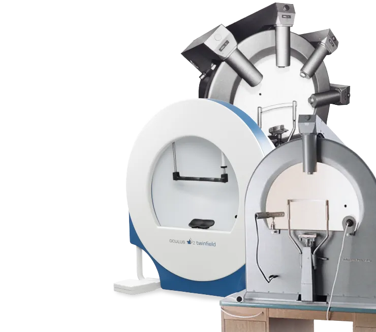
Diagram and Functions
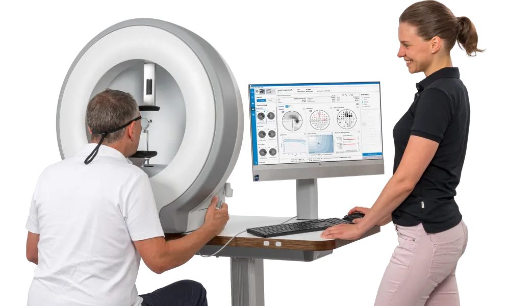
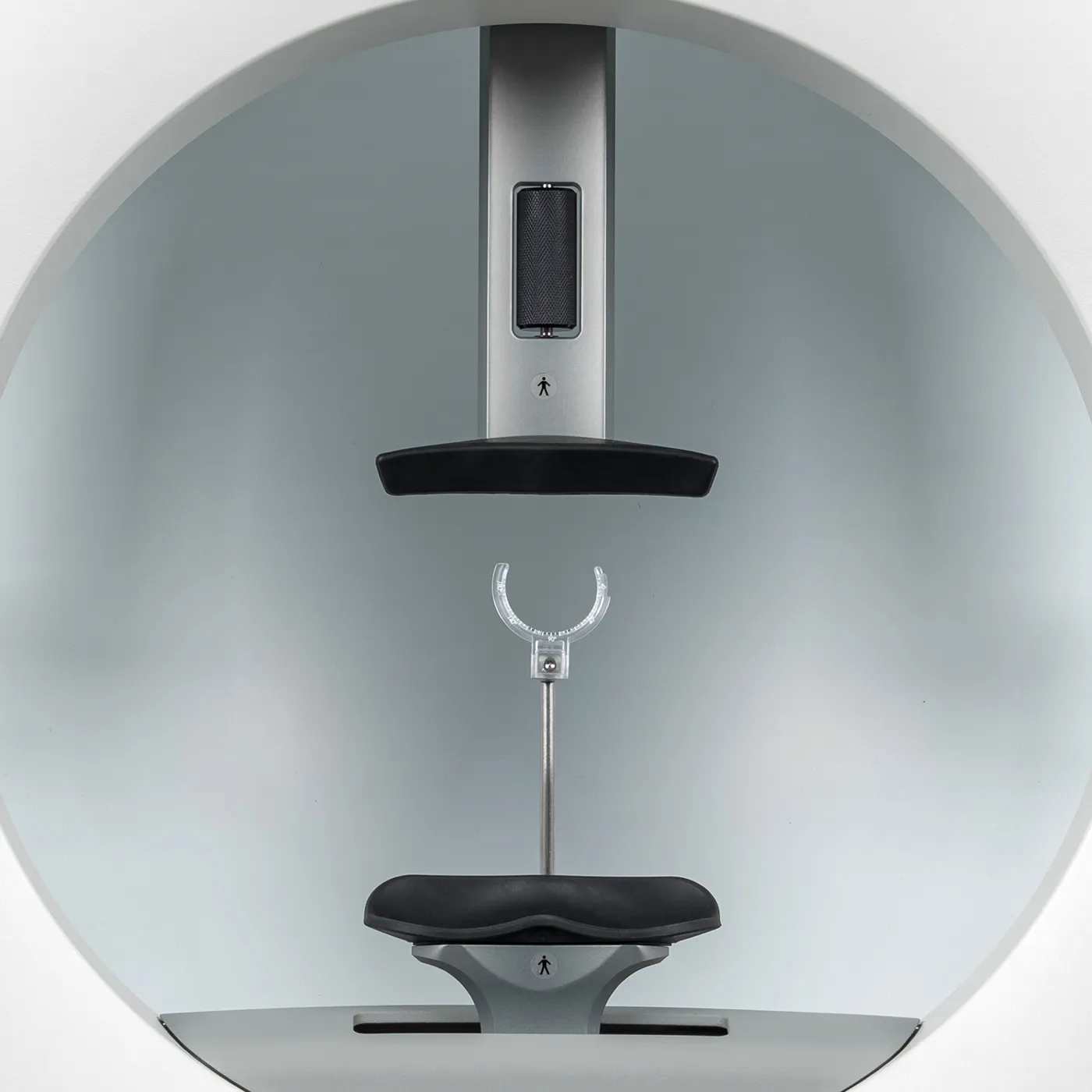
Improved ergonomics
The chin rest adjusts automatically, making it easier to position patients.
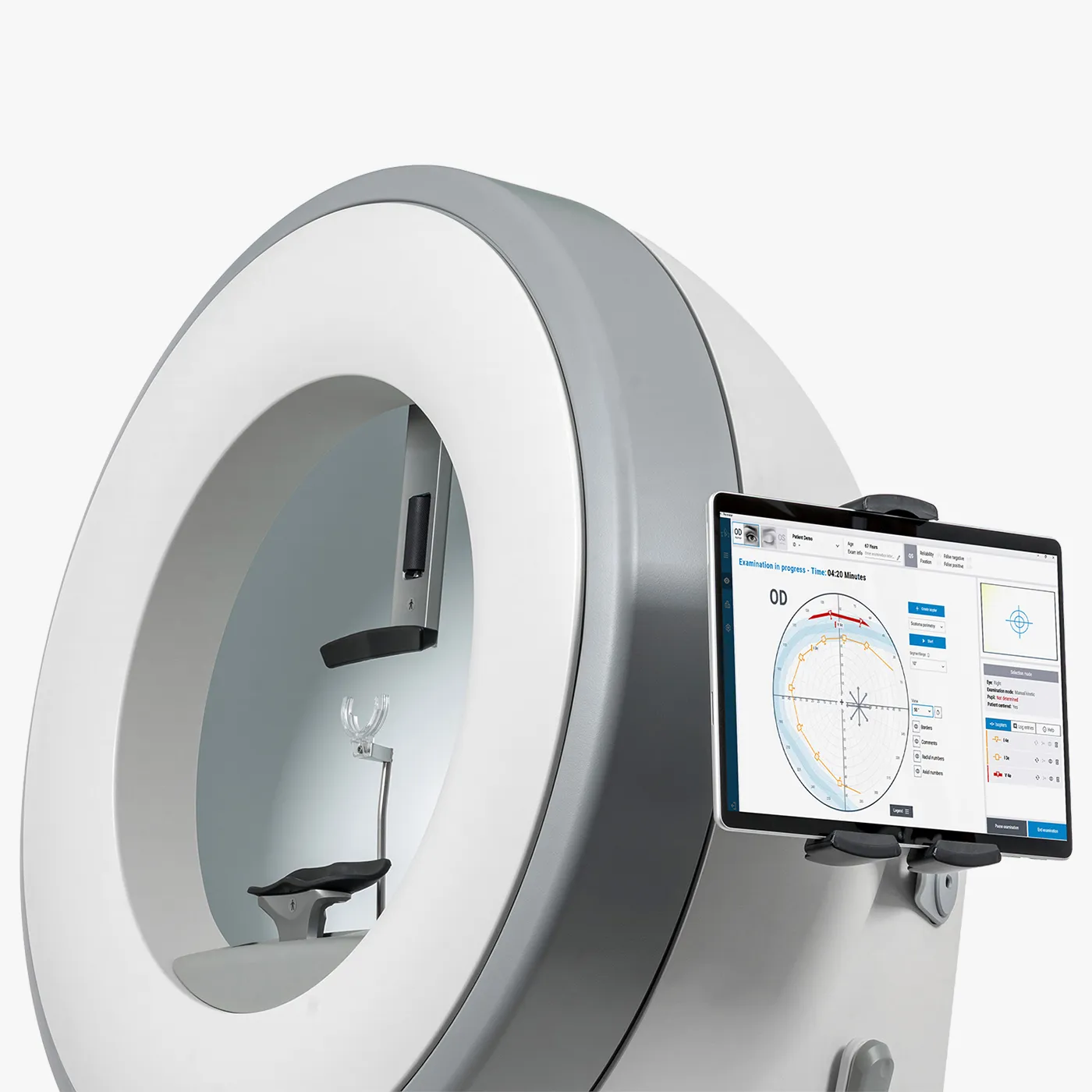
Tablet mount
An optional tablet mount is available.
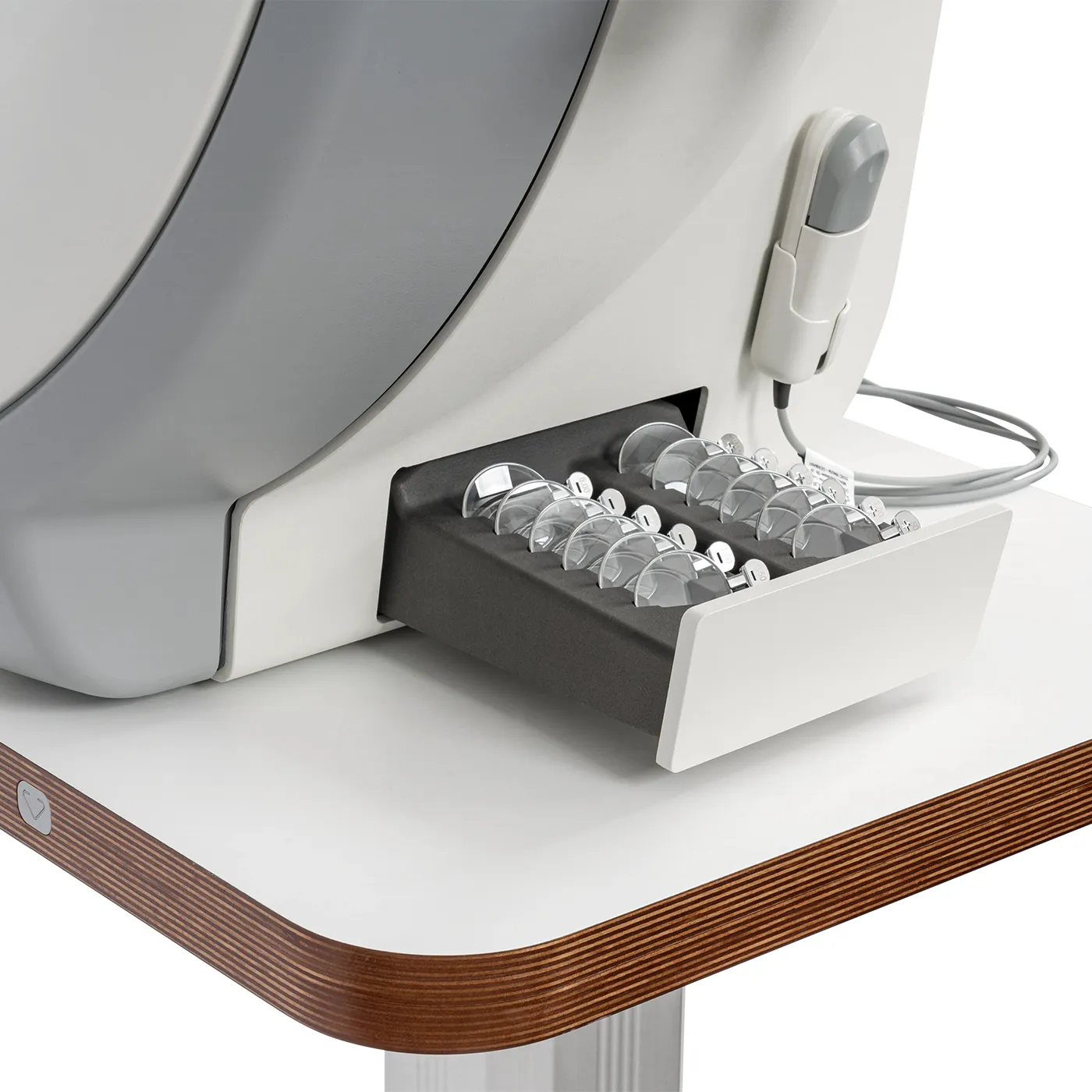
Thoughtful details
A built-in drawer stores slim rim trial lenses (full-aperture lenses), keeping them protected and within reach. The patient response button stores directly on the device.
Save time with audio instruction
Recorded audio instructions prepare patients before the test begins. With several languages to choose from, you can save time and help reduce language barriers in everyday practice.
Kinetic perimetry
Maximum flexibility
Kinetic perimetry uses moving light stimuli to measure the visual field. During the test, the patient focuses on a central fixation point while the examiner moves the light stimulus from the periphery to the center at a steady speed. The patient responds as soon as they see the stimulus. This maps lines of equal light sensitivity, known as isopters. The method can also define the outer limits of the visual field. It is particularly useful for disability evaluations and neurological assessments. Manual or assisted control gives you flexibility in how each test is performed.
Manual kinetic perimetry
Manual positioning and movement of the stimulus allow true Goldmann-style manual kinetic testing. All Goldmann stimulus sizes (I–V) are available.
Assisted manual kinetic perimetry
With assisted kinetic testing, you define the path of the stimulus, and the software moves it at a constant speed, independent of the examiner. This is designed to increase the reproducibility of the findings.
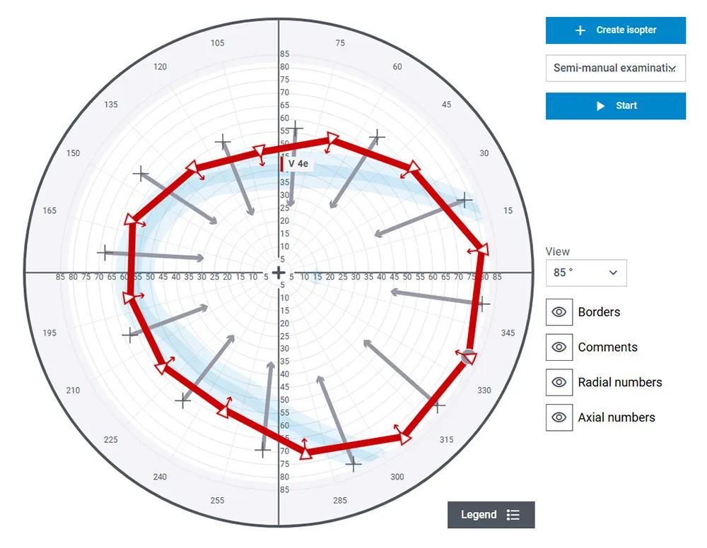
Application of assisted manual kinetic perimetry: after the V4e isopter is measured, segments are set for the I3e stimulus.
Scotoma mapping
Scotoma mapping supports repeated, systematic testing of visual field defects, such as targeted scanning of the blind spot.
Fully automated kinetic testing
Fully automated kinetic testing moves the stimulus along the meridians without manual control. It can be combined with static testing to perform quick screening of the entire visual field.
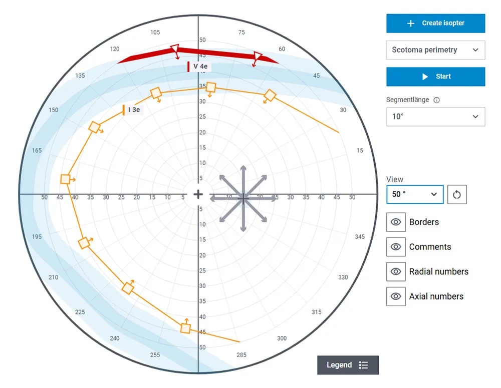
Scotoma mapping: the blind spot is scanned in eight directions using the I4e stimulus.
Glaucoma in focus
OCULUS threshold tests
For routine visual field testing in glaucoma patients, Twinfield® 3 offers a wide range of test programs with threshold strategies. Threshold strategies provide a complete picture of the visual field. The numerical sensitivity values can be evaluated statistically and used for quantitative progression monitoring.
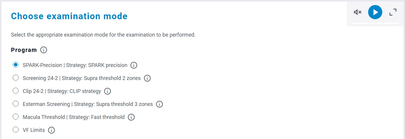
For a smooth workflow in everyday practice, the most frequently used test programs can be saved in a favorites list for quick selection.
SPARK – precision and speed in one strategy
The SPARK threshold strategy was developed specifically for glaucoma testing. It draws on the physiological relationships of photoreceptors along a retinal nerve fiber bundle to measure sensitivity values in the central visual field.
The visual field is measured in four phases in a maximum of three minutes per eye. Preliminary results are available in about 40 seconds.
Due to the short examination time and repeatability of the results, the SPARK threshold strategy is ideal for quantitative monitoring of glaucoma progression.
Other test strategies
The CLIP strategy (Continuous Light Increment Perimetry) is a very fast threshold strategy. The stimulus brightness increases continuously until the patient responds, which helps keep test time short.
OCULUS Fast Threshold is a widely used strategy with a shorter test time than the classic 4/2 threshold strategy.
Suprathreshold strategies offer shorter test times. The OCULUS Class Strategy assigns local sensitivity values to several classes, while the 2-zone and 3-zone strategies provide a simplified overview of the visual field.
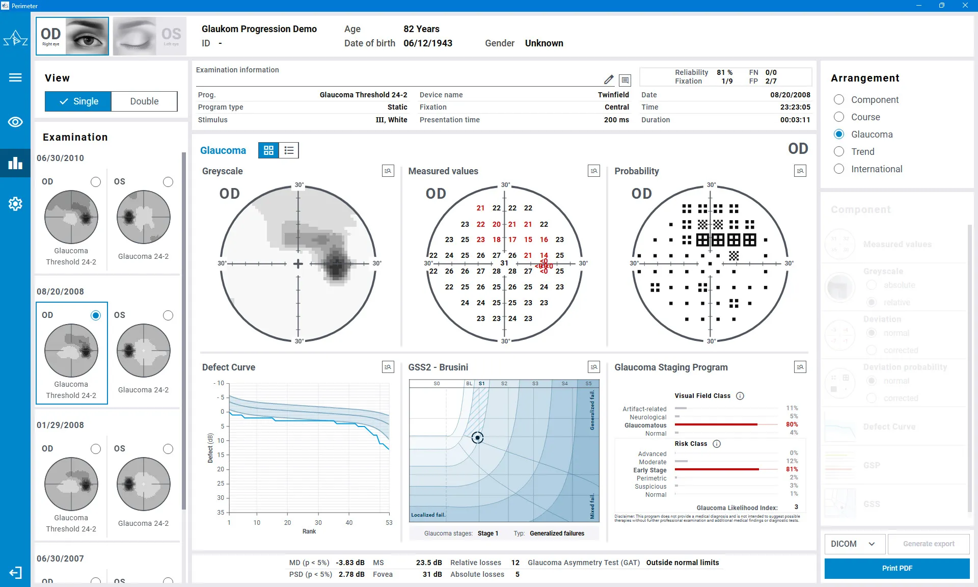
The glaucoma display:
A clear overview at a glance
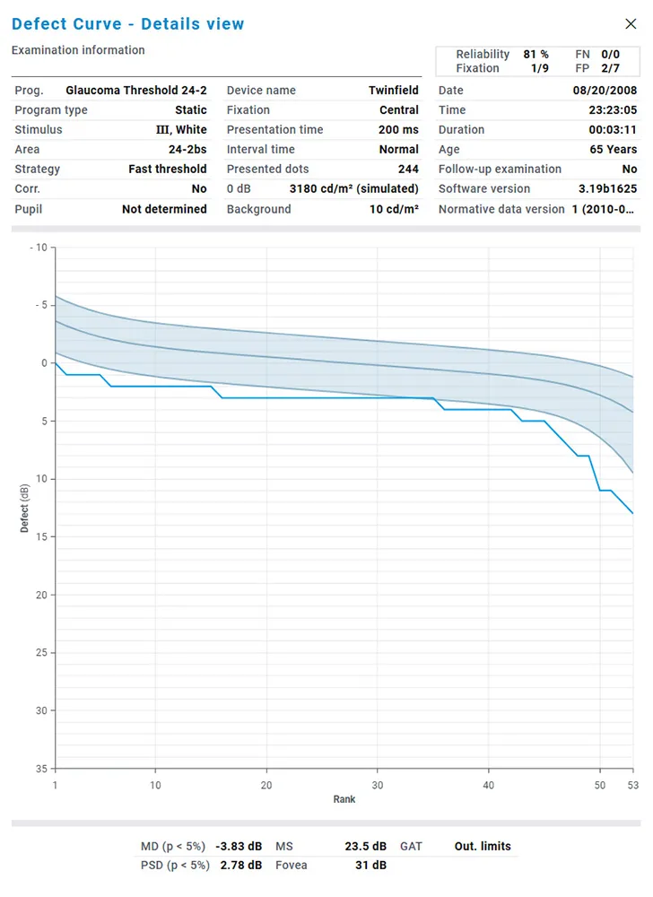
Cumulative defect curve
The cumulative defect curve is an alternative graphical representation of deviation values. It provides an overview of the entire visual field and helps differentiate local and diffuse defects.
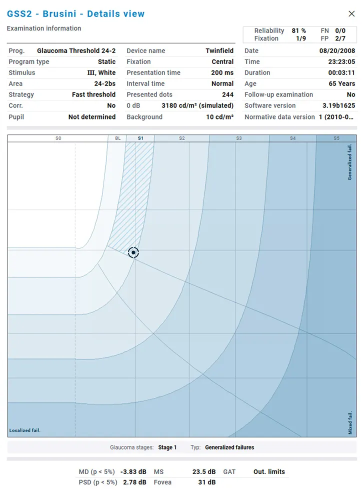
Intuitive classification of defects:
the Glaucoma Staging System (GSS 2)
The Glaucoma Staging System graphically classifies test results based on perimetric indices such as mean deviation (MD) and pattern standard deviation (PSD).
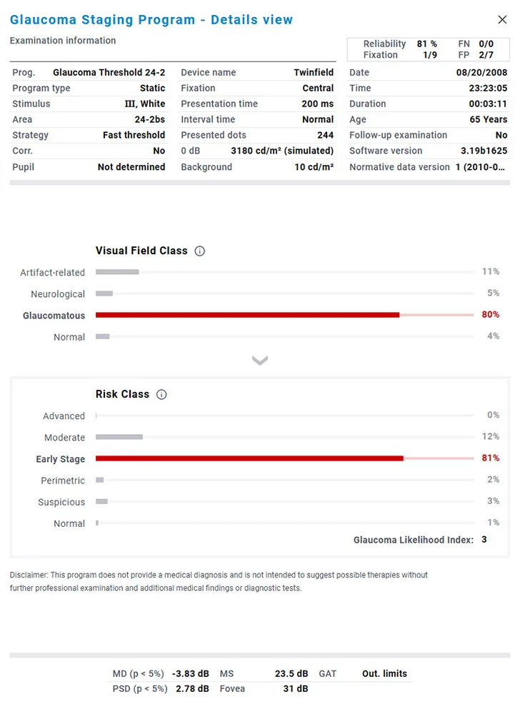
Evaluating risks:
Glaucoma Staging Program (GSP)
This evaluation module provides valuable support in the early detection of glaucoma and the assessment of findings. The GSP classification has been optimized to best correspond to the assessments of glaucoma experts. In addition to classifying the results, GSP also helps to assess the risks of different stages of glaucoma based on the visual field findings.
Quantitative progression analysis
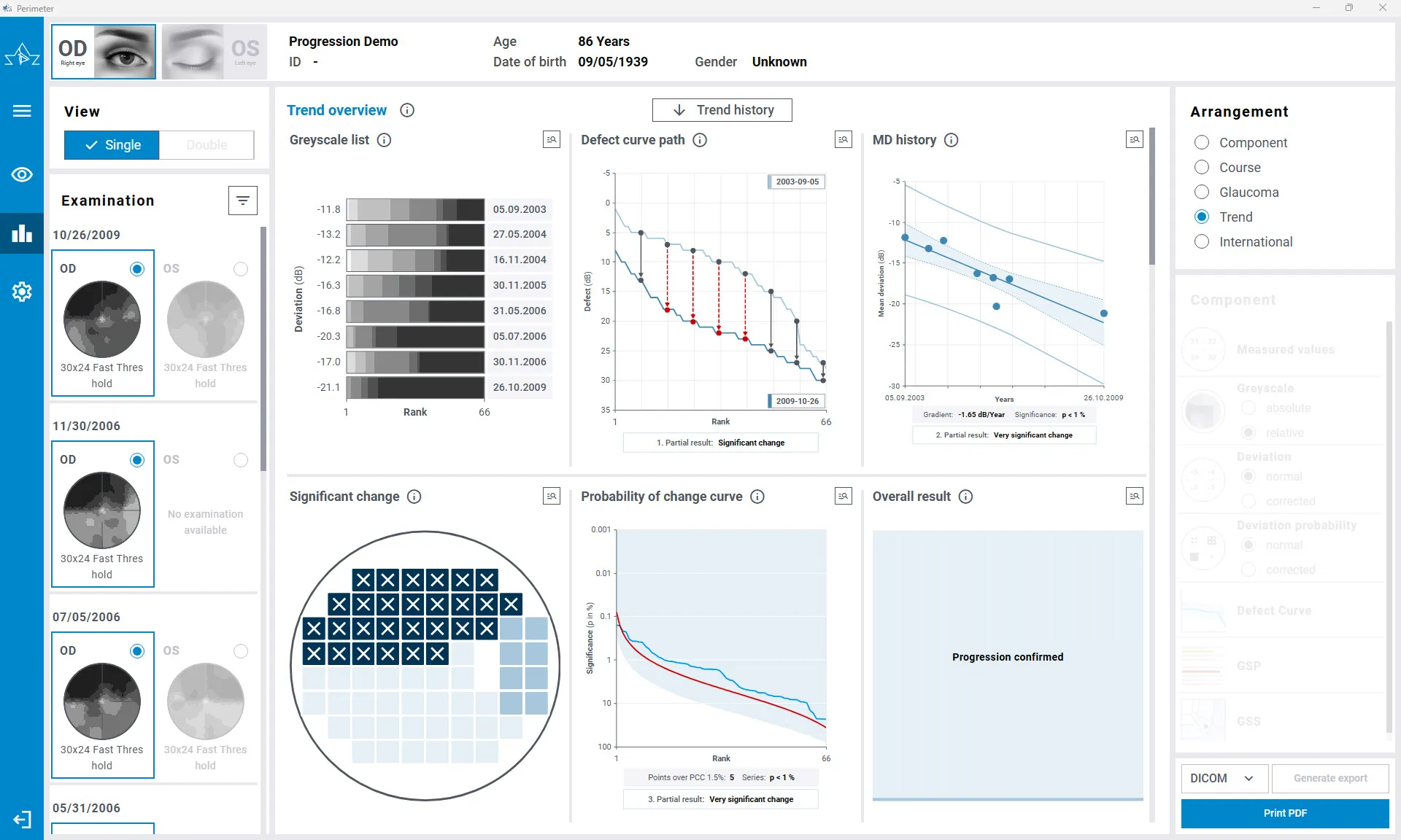
Visual field changes are assessed quantitatively by analyzing multiple parameters at once. The progression analysis is based on the Threshold Noiseless Trend (TNT) method, which evaluates the statistical significance of changes and quantifies progression as the rate of MD change per year (dB/year).
You may also be interested in these products
Smartfield
Perimetry
The OCULUS Smartfield is a compact visual field device purposefully optimized for monitoring functional impairment in glaucoma.
Easyfield®
Smallest Full-Fledged Perimeter on the Market
Enjoy combined visual field screening and comprehensive perimetry in one compact, lightweight device with an enclosed bowl design that enables practices to conduct exams in a lit room.
Centerfield® 2
Perimeter
The Centerfield® 2 Perimeter has proven itself to be an invaluable instrument in the Occupational Health area.
Technical Data
Static Perimetry
| Programs | Glaucoma, macula, neurological, static-kinetic screening and user-defined tests |
| Test Patterns | Physiological patterns (Area 1-8), orthogonal patterns (30-2, 24-2, 30×24, 10-2), Esterman, user-defined patterns |
| Strategies | Threshold Strategies: OCULUS Fast Threshold, Full Threshold (4/2) Optional: SPARK strategy, CLIP strategy Suprathreshold strategies: OCULUS Class Strategy, 2-zone, 3-zone, defect quantification |
| Speed | Adaptive, fast, normal, slow, user-defined |
| Fixation Control | Through central threshold, Heijl-Krakau (using the blind spot), live video image |
| Perimeter Bowl Radius | 300 mm (11.8 in) |
| Max. Eccentricity | 90° (full field) |
| Stimulus Sizes / Stimulus Colour | Goldmann I-V / white, blue, red |
| Stimulus Duratio | 200 ms, user-defined |
| Stimulus Luminance Range / Steps | 0 – 3,180 cd/m2 (0 – 10 000 asb) / 1 dB |
| Background Luminance / Background Colour | 10 cd/m2 / white | 100 cd/m2 / yellow |
| Optional reports | Glaucoma Staging Program (GSP), Threshold Noiseless Trend (TNT) progression report |
Kinetic Perimetry
| Measurement Methods | Automatic: Examination along meridians with freely selectable distances, Optional: Goldmann manual kinetic testing including: – Computer mouse freely moveable stimulus – Assisted kinetic testing along manually defined path – Scotoma mapping |
| Stimulus Speed | 2° / s (Goldmann standard) or user-defined |
Technical data
| Patient positioning | Motorized chin rest synchronized with adjustable forehead rest |
| Device dimensions (W x D x H) | 748 x 592 x 787 mm (29.4 x 23.3 x 31.0 in) – without table |
| Weight | 29 kg (64 lb) (without table and computer components) |
| Max. power consumption | 45 W (without table and computer components) |
| Voltage / Frequency | 100–240 V AC / 50–60 Hz |
| Recommended computer configuration | Intel® Core™ i5, 500 GB SSD, 8 GB RAM, Windows® 11, Intel® HD Graphics |
| Recommended screen resolution | 1920 x 1080 pixels (Full HD) |
| Interface | USB-A, USB-C |
| Software | Device control, patient management, backup and printing software (Windows®), integrated network operation, easy EMR integration, DICOM (optional) |
Contact us!
Request a quote or contact us if you need further information.
Request a quoteContact requestCall us
Questions?
Get in touch with us.
Tel. +1 888-284-8004