A picture is worth a thousand words
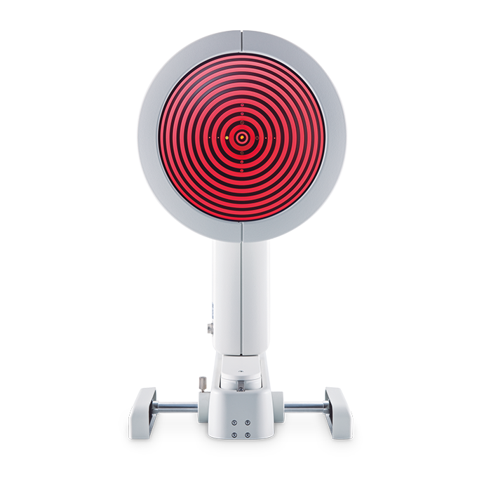
Gold standard corneal topography – that’s what the Keratograph® 4 is all about.
It ensures reliability when it comes to taking measurements, providing consultation and fitting contact lenses. The Keratograph® accurate findings are something you can count on.
The integrated keratometer and automatic measurement activation guarantee perfect reproducibility.
In this way the Keratograph® 4 also meets highest clinical standards for such procedures as tear film assessment and qualitative cornea analysis. It stands out by virtue of its versatility.
The keratometer: integrated in all OCULUS topography devices
The benefits of the integrated keratometer are crucial for opticians’ and optometrists’ everyday practice:
- measurements of actual corneal curvature
- exact measurements for perfect correction of contact lenses
- intuitive and simple to use
- can be processed quickly
- high-resolution images ensure clear results for customers
Highlights

A picture is worth a thousand words
Use the Keratograph® 4 as a marketing tool and incorporate it actively into your consultations. With the Keratograph® 4 software you can show images which your clients/patients have never seen before.

Contact Lens Fitting and Fluorescein Image Simulation
Contact lenses are recommended on an individual basis and displayed in a list. In order to avoid taking more steps than necessary when fitting contact lenses, the fluorescein image can be simulated beforehand.
The contact lens can be rotated and moved around. Fluorescein image simulation is adjusted automatically. The integrated and expandable database contains all customary types of contact lenses and is updated on a regular basis. The user can determine the order in which contact lens manufacturers appear.

OxiMap® – Visualizing the Oxygen Transmissibility
Professional patient consultation
The cornea needs oxygen and a good oxygen supply is fundamental for the comfort of a contact lens wearer. New materials used for soft contact lenses offer excellent oxygen transmissibility.
This can be shown with the new OCULUS OxiMap® display. You can easily show these color maps to your patients and help them choose better contact lenses.

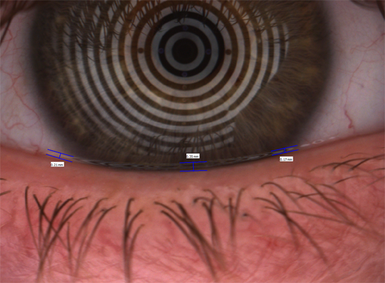
TF-Scan Illuminates the Condition of the Tear Film
Use this software to show patients the quality and quantity of their tear film through non-invasive measurements of the tear film breakup time as well as the tear film amount. Improve their understanding and trust in your assessment of their dry eye diagnosis.
Software
The heart of the Keratograph® 4
TF-Scan Makes the Tear Film Visible
Tear Film Quality (NIKBUT)
Tear Film Quantity (tear meniscus)
Different Ways of Determining Pupil Reaction:
Using the “Pupillometry” option
Optional Imaging-Software*
Essential for:
How Much Oxygen Really Reaches the Cornea?
Plain and Comprehensive Visualization Assures Patient Loyalty!
Compare Different Soft Contact Lenses!
In cases of dry eye patients and contact lens wearers, the tear film should be examined carefully. Only an intact tear film guarantees contact lens wearing comfort! The Keratograph® 4 measures the tear film breakup time non-invasively (quality assessment). You can show your patient the individual tear film quality using the color maps. In addition, you can take another non-invasive measurement to determine the amount of tear film (tear film quantity).
- Examination of the pupil reaction both with and without glare
- Examination using two different glare stimulus powers
- Clear presentation of the results in graphic form: pupil changes over a period of time; minimum, maximum and mean pupil diameter, incl. standard deviation
- Comparison views possible
* Optional software for Keratograph® 2007 April model and upwards
- Demonstration of the fit and mobility of contact lenses
- Assessment of the static fluo-image
- Assessment of the fit of the contact lenses with different pupil diameters
- Side-by-side comparison of fluo-image simulations to real-time fluo-images
- Choice of the final contact lense
- Expertise and customer long term relationship
Until now, only the oxygen transmissibility values for the center of a contact lens with -3.0 D were available. The OxiMap® shows the oxygen transmissibility depending on the lens material and the lens thickness. The OxiMap® is available for the most frequently sold spherical soft contact lenses. This impressive tool assists you in helping your patients select the most suitable contact lens.
OxiMap® was developed in close cooperation with JENVIS Research and the University of Applied Sciences in Jena.


Title
Description
FAQs
Specifications
Technical Data
| Measuring range | 0.1 – 1.5 in, 9 – 99 D |
| Accuracy | ± 0.1 D |
| Reproducibility | ± 0.1 D |
| Number of rings | 22 |
| Working distance | 80 mm |
| Number of measuring points | 22,000 |
| Camera | digital CCD camera |
| Source of illumination | placido illumination: red 650 nm imaging illumination: blue 465 nm (UV-free) pupillometer illumination: infrared 880 nm |
| Dimensions (W x D x H) | 10.8 x 12.6 to 15.7 x 19.3 to 20.4 in |
| Weight (Measuring head) | 5.1 lbs |
| Weight (with base) | 11.7 lbs |
| Power Consumption max. | 30 W |
| Voltage | 100 – 240 V AC |
| Frequency | 50 – 60 Hz |
| Minimum PC requirements | Intel® Core™ i5, 1 TB HDD, 8 GB RAM, Windows® 7 – Windows® 10 |
| Recommended screen resolution | 1920 x 1200 pixel |
Contact us!
Request a quote or contact us if you need further information.
Request a quoteContact requestHotline
Questions?
Get in touch with us.
Tel. +1 888 284-8004
Choose your topographer
OCULUS gives you the choice. What do you need for topography in your everyday practice?
Keratograph® 5M, Keratograph® 4 or Easygraph: Whichever device you choose, OCULUS quality is assured.
 |
|
 |
|
| Keratograph® 5M | Keratograph® 4 | Easygraph | |
| Topography | |||
| Overview Presentation | ✓ | ✓ | ✓ |
| Large Color Map | ✓ | ✓ | ✓ |
| 4 Color Maps Selectable | ✓ | ✓ | ✓ |
| Camera Image | ✓ | ✓ | ✓ |
| 3D Cornea | ✓ | ✓ | ✓ |
| Fourier Analysis and Zernike Analysis | ✓ | ✓ | ✓ |
| Topographic Keratoconus Screening | ✓ | ✓ | ✓ |
| Elevation Map | ✓ | ✓ | ✓ |
| Corneal asphericity | ✓ | ✓ | ✓ |
| Lens Fitting | ✓ | ✓ | ✓ |
| Compare examinations | ✓ | ✓ | ✓ |
| Measurement contact lens back surface | ✓ | – | – |
| Imaging | |||
| Image and video documentation with fluo images | ✓ | ⃘ | ✓ |
| Near-Portion Height Measurement | ✓ | ✓ | – |
| Palpebral Angle Measurement | ✓ | ✓ | – |
| Manual Recording Mode | ✓ | ✓ | – |
| Crystal TEAR Report | |||
| Crystal TEAR Report (Find out the cause of dry eye disease quickly and reliably) | ⃘ | – | – |
| TF-Scan (Evaluation of lipid layer and tear film dynamics, measurement of tear meniscus height and non-invasive tear film break-up time (NIKBUT)) | ⃘ | ⃘ | – |
| R-Scan (Automatic classification of bulbar and limbal redness) | ⃘ | – | – |
| Meibo-Scan (Meibography of upper and lower eyelid) | ⃘ | – | – |
| Pupillometry | |||
| Examination of pupillary response using the pupillometer, asymmetry test and manual measuring mode | ⃘ | ⃘ | – |
| Oximap® | |||
| Raphic representation of the oxygen transmissibility (Dk/t) of soft contact lenses | ⃘ | ⃘ | ⃘ |
| Technical Data | |||
| Measuring range | 0.1 – 1.5 in, 9 – 99 D | 0.1 – 1.5 in, 9 – 99 D | 0.1 – 1.5 in, 9 – 99 D |
| Accuracy | ± 0.1 D | ± 0.1 D | ± 0.1 D |
| Reproducibility | ± 0.1 D | ± 0.1 D | ± 0.1 D |
| Number of rings | 22 | 22 | 22 |
| Working distance | 78 bis 100 mm | 80 mm | 40 mm |
| Number of evaluated data points | 22,000 | 22,000 | 22,000 |
| Camera | Digital CCD camera | Digital CCD camera | – |
| Dimensions (W x D x H) | 275 x 320 to 400 x 480 to 510 mm (10.8 x 12.6 to 15.7 x 18.9 to 20.2 in) |
275 x 320 to 400 x 490 to 517 mm (10.8 x 12.6 to 15.7 x 19.3 to 20.4 in) |
119 x 102 x 216 mm (4.7 x 4.0 x 8.5 in) |
| Weight | 7.1 lbs (Measuring head) / 13.5 lbs (with base) | 5.1 lbs (Measuring head) / 11.7 lbs (with base) | 1.6 lbs |
| Details | Details |
Legend: ✓ = available * – = not available * ⃘ = optionally available against surcharge