 Workstations / Ophthalmic tables
 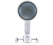 Topography
 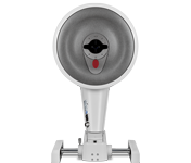 Tomography
 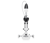 Slit Lamp / Documentation
  Refraction Equipment
  Myopia Management
  Perimetry
  Tonometer
 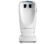 Visual Test Equipment
 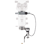 Vitreoretinal Surgery
  Refraction
  Vision Test Types
 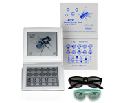 Stereo Tests
  Eye Test Units
  Orthoptics / Pleoptics
  Other Products
 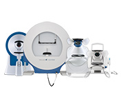 Perimetry
 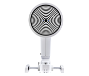 Dry Eye
 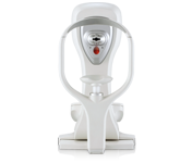 Corneal Biomechanics
 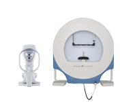 Glaucoma
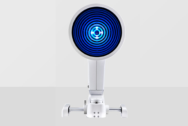
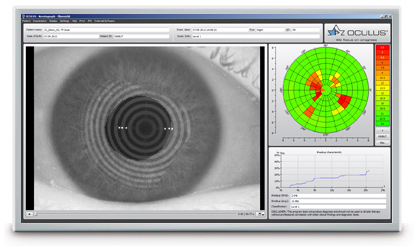
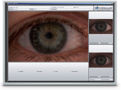
The OCULUS Keratograph 5M is an advanced corneal topographer with a built-in real keratometer and a colour camera optimized for external imaging. Unique features include examining the meibomian glands, non-invasive tear film break-up time and the tear meniscus height measurement and evaluating the lipid layer.

|
OCULUS Keratograph 5M |


With the Keratograph 5M, you can show your patients images they have never seen before. Gain patient trust by providing professional consultation during examinations and follow-ups.
Actively integrate the Keratograph into your consultation. Many easy to understand displays support you in communication and patient education. Use your Keratograph 5M as a marketing tool to make your services transparent!
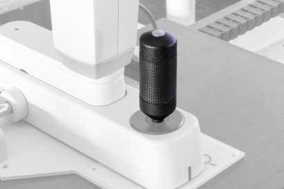
The new joystick for the Keratograph 5M allows you to capture images and video sequences directly from the device. No need to use the mouse: your hand just stays on the joystick when you capture images or videos.
The new joystick communicates via a smart Bluetooth interface. And it does away with the need for cumbersome power cabling. The joystick switches to sleep mode function when not in use to extend battery life.
Switching to the new joystick is quick and simple. Watch the video below to see how easy it is!
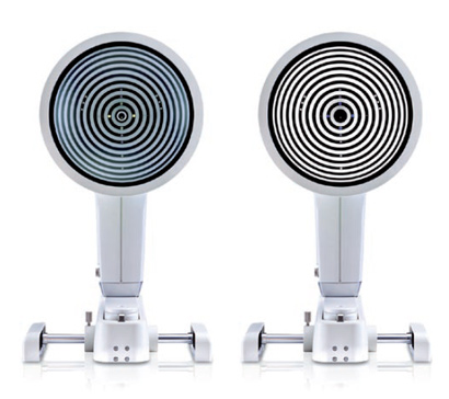
Thousands of measuring points are used to measure the whole surface of the cornea. A white ring illumination is used for this purpose. An infrared ring illumination is also provided for analysis of the tear film to prevent glare-related reflex secretion.
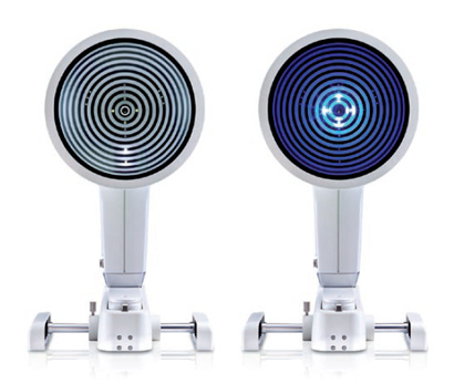
The perfect illumination has been integrated for every function of the Keratograph 5M: White diodes for the tear film dynamics, blue diodes for fluo-images, infrared diodes for Meibography.
 |
| Accuracy: | ± 0.1 D |
| Reproducibility | ± 0.1 D |
| Number of rings: | 22 |
| Working distance: | 78 - 100 mm |
| Number of evaluated data points | 22 000 |
| Camera: | Digital CCD camera |
| Light source: | Placido illumination: White Placido illumination: Infrared 880 nm Fluorescein illumination: Blue 465 nmMeibography: Infrared 840 nmTear film dynamics: White Pupillometer illumination: Infrared 880 nm |
| Dimensions (W x D x H): | 275 x 320 - 400 x 485 - 512 mm |
| Weight | Measuring head: 3.2 kg (7.1 lbs) With base: 6.1 kg (13.5 lbs) |
| Max. power consumption | 18 W |
| Voltage | 90 - 264 V AC |
| Frequency | 47 - 63 Hz |
| Recommended computer specifications | CPU Intel® Core i5-7600, 8 GB RAM, 1 TB, Windows® 10 Pro |
| Recommended screen resolution | 1920 x 1200 pixel |
In acc. with the Medical Device Directive 93/42/EEC

The OCULUS QM system is certified in accordance with ISO 13485 and (EU) 2017/745 (MDR).

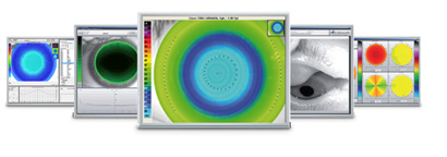
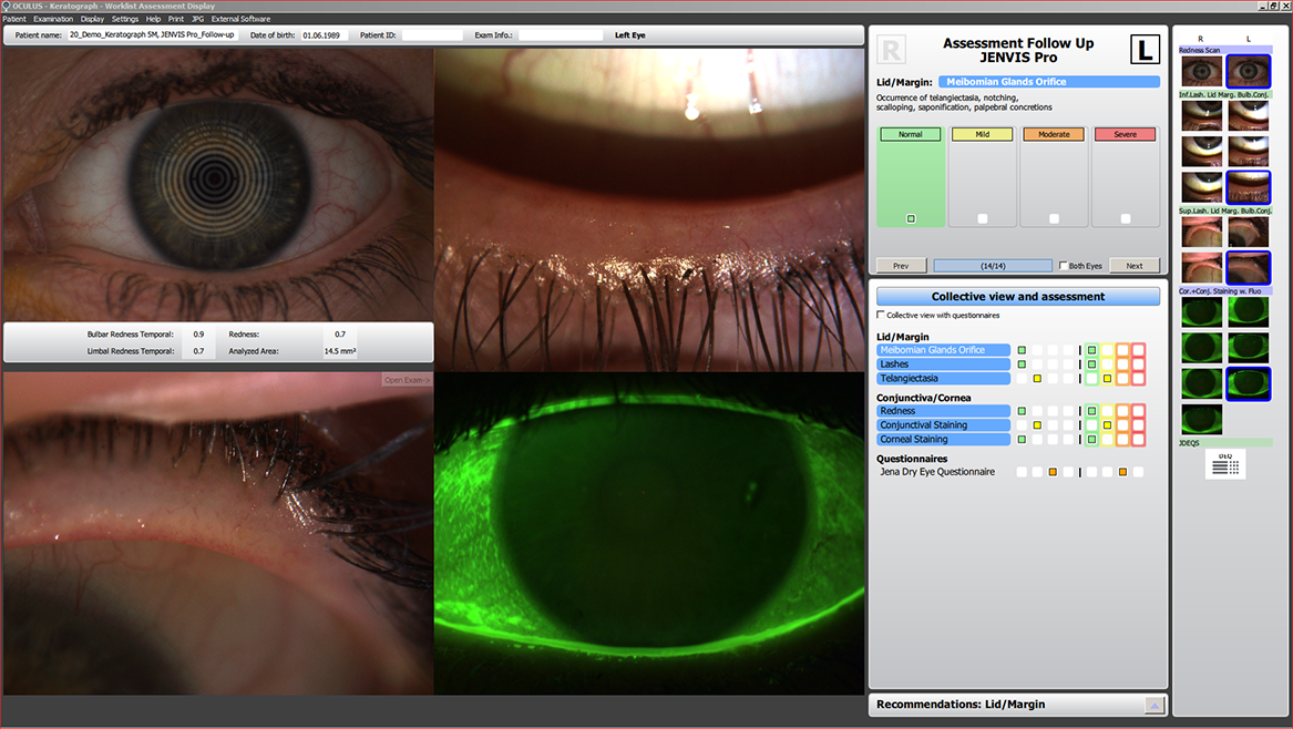
Find the cause of dry eye syndrome quickly and reliably. The new JENVIS Pro Dry Eye Report in the Keratograph 5M will help you as you go along. Perform a comprehensive screening, using the measuring results as a basis for diagnosing dry eye syndrome. All results are documented in accordance with the Medical Products Law and summarized for your patient in a neat and easily understandable printout.
Make use of all the advantages of the new JENVIS Pro Dry Eye Report in the Keratograph 5M: efficient screening, well-founded measurement results and greater patient loyalty.
Screening for dry eye syndrome should be a routine part of every refraction.
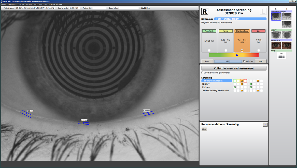
The four-part screening test quickly and accurately reveals abnormalities in tear film quantity and quality. This 5-minute screening should be carried out routinely before every refraction. The JENVIS Pro tear film screening routine includes measurement of the tear meniscus height, tear film break-up time, bulbar redness and a short questionnaire.
Make a quick and easy decision whether your patient has dry eye symptoms.
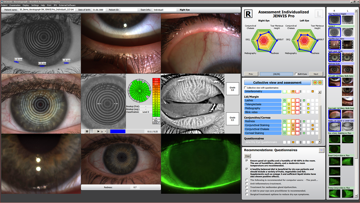
The JENVIS Pro Dry Eye Report provides you with a fail-safe test sequence covering all the assessment criteria required for a comprehensive analysis for dry eye syndrome. It provides you with useful hints and optimized, predefined settings to support you in your measurements, enabling you to perform a quick and efficient analysis for dry eye syndrome and document your anterior segment findings.
Get equipped to give your patients specific advice for easing their dry eye symptoms.
You want to know whether your treatment recommendations have been successful? Then use the follow-up function with its setting options for characterizing the initial cause. This will enable you successfully document improvements in your patients‘ tear film condition.
Regular check-ups will make you the go-to specialist for your patients.
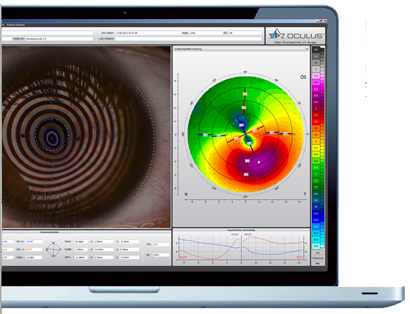
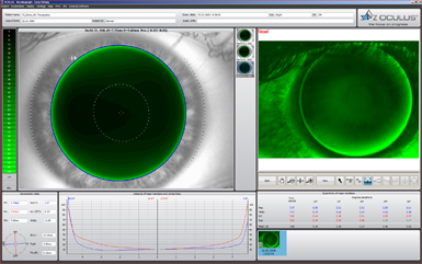
The tear film is assessed using either white or infrared illumination. The new high-resolution colour camera makes even the finest of structures visible. In addition to NIKBUT (Non-Invasive Keratograph Break-Up Time) and measurement of the meniscus tear, the TF-Scan can also make an assessment of the lipid layer and the tear film dynamics. Tear film analysis with the OCULUS Keratograph 5M is non-invasive and is conducted without any additional tools.
The tear film break-up time is measured non-invasively and fully automatically. The new infrared illumination is not visible to the human eye. This prevents glare during the examination. The TF-scan presents the results in a way that is easy for you and your customers to understand.
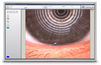
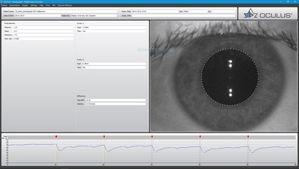
The height of the tear meniscus can be precisely measured with an integrated ruler and various magnification options, and its development along the edge of the bottom lid can be assessed. The results are saved to the patient file.
The interference colours of the lipid layer and their structure are made visible and can be recorded. The thickness of the lipid layer is assessed based on the structure and colour.
The video recording, with up to 32 frames per second, enables the observation of the tear film particle flow, from which conclusions regarding the viscosity of the tear film can be drawn.

Automatic classification of the bulbar redness. Conjunctival redness used to be assessed subjectively and depended on the examiner. The R-Scan is the first module that automatically and objectively documents and classifies the bulbar and limbal degree of redness. The R-Scan detects the blood vessels in the conjunctiva and evaluates the degree of redness. This automatic assessment spares you the time and effort of having to make manual comparisons using classification sheets.
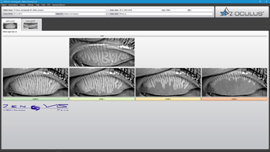
The multi-functionality of the Keratograph 5M easily and efficiently integrates difficult examinations such as meibography. The dysfunction of meibomian glands is the most frequent cause of dry eye. Morphological changes in the gland tissue are made visible using the Meibo-Scan and can be classified with the JENVIS Meibo Grading Scales.
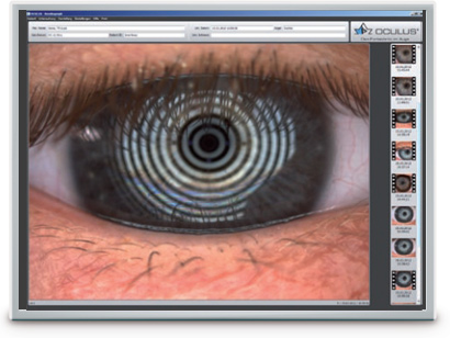
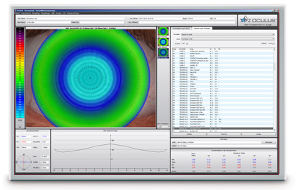
The Imaging Software is used to record video and image files. In addition to viewing the videos and single images by themselves, you can also compare the recordings with the simulated fluorescein-images of the measured eye.
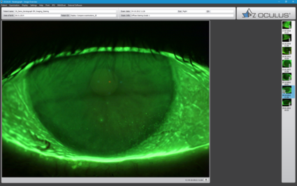
Result: Static fluorescein-images and videos can be recorded under slit lamp conditions!
The OxiMap® presents a colour map of the oxygen transmissibility of soft contact lenses based on the lens power, which is easy to understand – even for your customers!
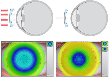
An intact tear film and a good supply of oxygen to the cornea are an absolute necessity for wearing contact lenses comfortably. The OxiMap® presents a colour map of the oxygen transmissibility of soft contact lenses based on the lens power – which even your customers will understand!
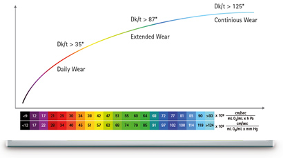
The oxygen transmissibility (Dk/t) depends on the material the contact lens is made of and on its thickness. In the past, only the manufacturers’ specifications about the oxygen transmissibility in the center of a -3,00 D contact lens were available. With the OxiMap®, you now get a graphic visualization of the Dk/t values over the whole area, based on the lens thickness.
The Dk/t values are colour-coded, whereby black represents an oxygen supply lower than that present in a closed eye. To preserve the integrity of the cornea when wearing contact lenses, minimum Dk/t values are recommended based on the length of time the contact lenses are to be worn. The OxiMap® colour-coding is based on these international recommendations.
The OxiMap® is individually adapted based on the lens power and supports you in your consultation with the patient and helps you to choose the most suitable contact lens. New materials used for soft contact lenses provide excellent oxygen transmissibility.
OxiMap® was developed in close cooperation with JENVIS Research and the University of Applied Sciences in Jena.
Author: Dr Jennifer Rayner, Optometrist, BAppSc(Optom), GradCertOcTher(UNSW)
Alleve Eye Clinic, Adelaide, Australia
In a perfect world, patients would present with a single, obvious cause for their dry eye and management would be simple, quick and effective. Yet there are often multiple factors that contribute to the presence of dry eye and they need to be identified as contributors for best management. A tool such as the OCULUS Keratograph 5M with specific JENVIS Pro Dry Eye Report software allows for advanced testing to understand the subtleties and complex nature of the ocular surface and tear-film.
Author: Dr. Keyur Patel, BSc(Hons) OD, DipTP(IP) DipGlauc DipSpV FAAO FCOptom FBCLA Tompkins, Knight & Son Optometrists, UK
More often than not, patients do not present with a text book case of a single issue. Usually, they have a combination of concerns that may or may not be relevant to each other. In the world of contact lenses, comfort and drop out are often related to a poor ocular surface, rather than the latest generation of contact lens products. This case report demonstrates how multi-tasking tools, such as the OCULUS Keratograph 5M, allow us to investigate multiple issues and concerns in a parallel stream allowing maximum efficiency and outcome.
Dry Eye Examination
Can the NIKBUT measurement be performed even in severe dry eye?
The NIKBUT measurement for dry eye with severe corneal epithelial damage is difficult. Even when the NIKBUT measurement is performed in such cases, the device mostly reports the results as “too short”.
Is the NIKBUT value the same as fluorescein tear film break-up time (TFBUT)?
The differences between fluorescein TFBUT and NIKBUT values should be noted, although positive correlation between TMH and Schirmer test values has been reported.
Why are white areas sometimes displayed in the colour-coded map of the NIKBUT display?
White areas are displayed if the patient blinks before 15 seconds after the measurement started. This does not affect the measurement in any way.
Does abnormal corneal shape i.e. keratoconus affect the NIKBUT measurement?
No, the software detects a basic image. In the case of keratoconus for example the strained Placido rings induced by the condition is noted initially by software and distortions from the original image is recorded and indicated as a break in the tear film. However, in cases with advanced keratoconus or severe irregular corneal astigmatism, it is hard to perform the measurement.
Does the nose shadow affect the NIKBUT measurement?
No, the area being analyzed during the NIKBUT measurement usually doesn’t coincide with the area affected by the nose shadow.
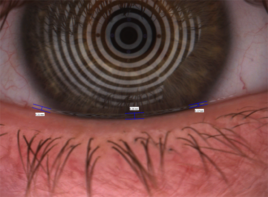
What is important when measuring tear meniscus height (TMH)?
At least three measurement points (nasal and temporal limbus, pupil center) should be selected. To get the right value the ruler has to be placed on the lid margin and measure perpendicular up to the last reflex of the meniscus. Please see image as reference. The correct measurment is 0.22 mm for this example.
Are the meibomian glands graded objectively by the software?
No, however grading scales called the JENVIS Grading Scales for meibomian glands have been included in the software. A sliding bar tool allows for direct comparison of patient results to the grading scale, making meibo grading very easy with the Keratograph 5M.
In some cases, R-Scan evaluating bulbar redness cannot detect the conjunctival area correctly.
In eyes with small lid aperture or eyes with ptosis, (commonly seen in the older Asian population), the device cannot detect the conjunctival area correctly.
What are the particles being observed during the tear film dynamic examination?
The particles are a combination of eroding corneal epithelial cells, tarsal conjunctival cells and small air-particles.
The JENVIS Pro Dry Eye Report is a comprehensive dry eye report. Does this report diagnose the condition of the eye?
The purpose of the JENVIS Pro Dry Eye Report is to provide the clinician with various dry eye related examinations, improving workflow, aiding in the diagnosis and educating the patient. The clinician uses the results from the JENVIS Pro Dry Eye Report to formulate the diagnosis. Treatment options are included in the software, however these are chosen at the discretion from the clinician. Different treatment suggestions can be manually added.
Various tests are included in the JENVIS Pro Dry Eye Report, does all of the tests need to be performed on every patient?
No. The report is set up to allow clinicians to individualize the screening procedure if they so require. A screening option, consisting only 4 procedures, is also included. This is a good tool to screen every patient entering the clinic. If a problem is detected, more detailed examination with an individualized report can follow.
In which sequence should dry eye tests be performed?
It is important to start from least invasive followed by more invasive tests. The workflow in the JENVIS Pro Dry Eye Report is set up to accommodate this principle.
Can data from the Keratograph be exported for research?
Yes, data from the NIKBUT, R-Scan and all keratometric data can be exported to csv files.
Topography/Contact lens fitting
The Keratograph has 22 Placido rings to perform a measurement. Does this generate sufficient information about the cornea?
Yes, with the 22 Placido disc 22.000 real measured data points are generated after each measurement. None of these are extrapolated measurements.
Does the Keratograph measure simKs?
Yes, a real keratometer is included in the hardware measuring simKs of the anterior surface.
Does the Keratograph measure the posterior cornea?
No, the Keratograph is a Placido topographer. Placido discs are projected on the tear film, the software analyzes the resulting images and measurement data of the anterior cornea is generated. No, a Pacido topographer can measure the posterior cornea on its own.
How does the Keratograph aid in keratoconus diagnosis?
Topographic indices for keratoconus screening are incorporated in the software, as well as Fourier Analysis. Objective inspection of the sagittal map is another tool to aid with diagnosis.
The OxiMap® displays oxygen transmissibility of a soft contact lens. Can any lens on the patient’s eye be measured?
No, lenses cannot be individually measured. Well-known soft lenses were analyzed and the information is programmed into the software. The role of the OxiMap® is to educate patient’s on the importance of using lenses with high oxygen transmissibility, showing them the benefits in an easy to understand image. Furthermore the OxiMap® gives information about the oxygen transmissibility over the entire surface of the lens, as well as differences between prescriptions.
Data of various lenses are included in the Contact Lens Fitting display. Can data for other lenses be imported too?
Yes, data from any lens can be manually input if the parameters are known.

Product Categories


|
The OCULUS QM system is certified in accordance with ISO 13485 and (EU) 2017/745 (MDR). |